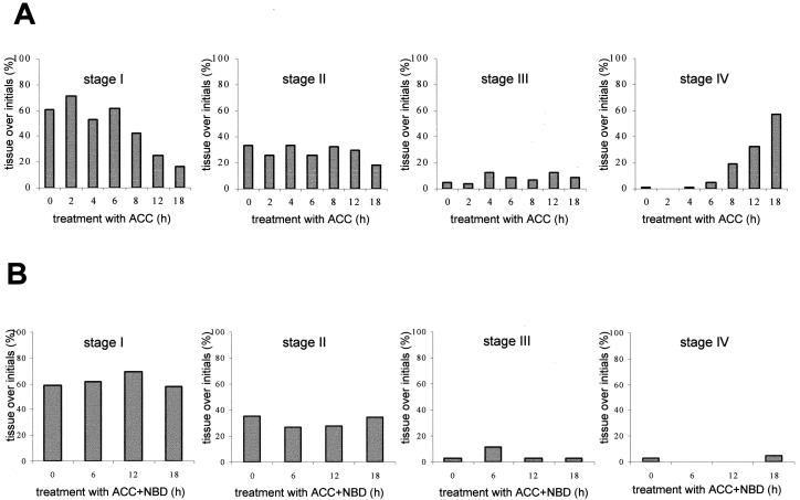
Figure 4.
Degree of epidermal Evans blue staining above root primordia at the third node of isolated stem sections. A, Stem sections were treated with 10 mm ACC for the times indicated. Results are averages of at least 15 stem sections analyzed in three independent experiments. Each stem section contained 12 to 15 adventitious root primordia. B, Stem sections were treated with 10 mm ACC and 50 μL/L NBD for the times indicated. The staining patterns are given as percentages of stage I to stage IV as described in Figure 3A. Results are averages of at least five stem sections analyzed.

