Figure 1.

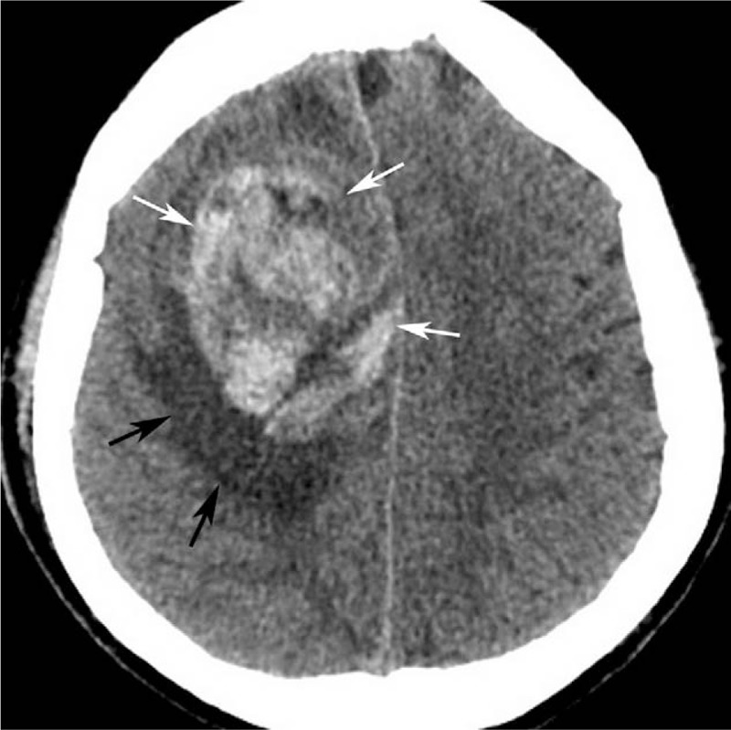
Computed tomography (CT) plain scan showed a round, heterogeneous hyper-attenuating mass (white arrow) with peripheral edema (black arrow) in the right frontal lobe. CT = computed tomography.

Computed tomography (CT) plain scan showed a round, heterogeneous hyper-attenuating mass (white arrow) with peripheral edema (black arrow) in the right frontal lobe. CT = computed tomography.