
Figure 2.
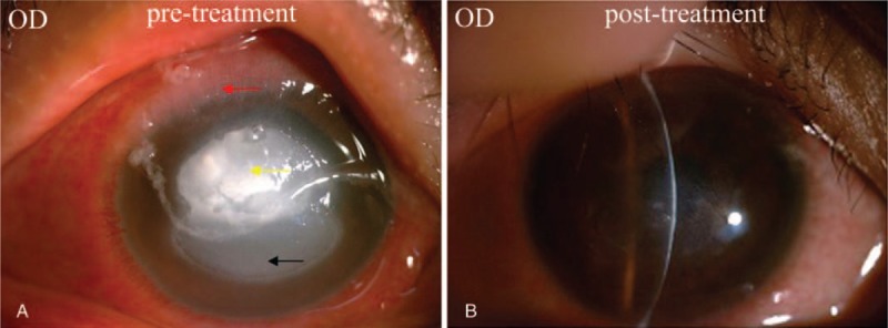
(A) Diffuse superficial conjunctival congestion, gelatinous hyperplasia at the superior limbus (red arrow), shield ulcer in the inferior cornea (black arrow). The superior of the shield ulcer showed dense multifocal anterior stromal infiltrate accompanied with necrotic tissue (yellow arrow). (B) Nebulomacular corneal scar postresolution.