
Figure 3.
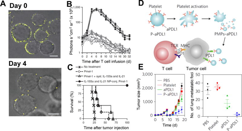
Cell membrane conjugation on T cells (A-C) and platelets (D-E) for enhanced cancer immunotherapy. Liposomes encapsulating cytokines for T cell proliferation can be attached on T cells to overcome their inhibited proliferation in tumors. (A) Confocal images of T cells conjugated with fluorescently labeled liposomes on day 0 and day 4 after stimulation. Scale bars, 2 μm (B) Bioluminescence intensity of T cells variation with time. The T cells expressed firefly luciferase, and the bioluminescence intensity was an indicator of T cell numbers in the mice. Each curve represents one mouse. (C) Survival curves of B16 melanoma-bearing mice after T cell therapy. Immune check inhibitor can be delivered by platelets to residual tumors after surgical removal of primary tumors. (D) Illustration of aPDL1 conjugated on platelets for blocking PDL1 on tumor cells to enhance T cell antitumor function. (E) Tumor growth curves of mice with an incomplete surgical resection of mouse melanoma tumors (left). Numbers of lung metastatic foci from mice treated with PDL1 antibody-modified platelets (right). (Reproduced with permission from Ref 37, Copyright© 2010, Nature Publishing Group; and Ref 54, Copyright© 2017, Nature Publishing Group)