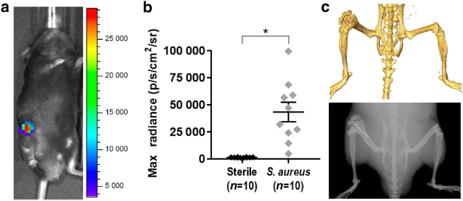
Fig. 1.
In vivo bioluminescent imaging in a mouse model of PJI. A titanium Kirschner-wire was surgically placed in the mouse femur with or without an inoculum of a bioluminescent S. aureus strain in the knee joint prior to closure. a Representative in vivo bioluminescent signals at the S. aureus-infected postoperative site. b Maximum radiance values for individuals (mean ± S.E.M.) with sterile control implants or S. aureus-infected postoperative distal femur sites at a representative time point (1 400 ± 90 vs 43 000 ± 9 000, respectively). c CT (top) and X-ray (bottom) imaging demonstrating the location of the Kirschner-wire (false-colored pink in the CT image) in the distal femur. *P < 0.000 6

