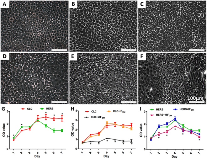
Figure 2.
Morphological observation and CCK-8 analysis of CLC and HERS cells after induction by IFCM and MFCM. Compared with non-induced CLC (A) and HERS cells (D), CLC induced with IFCM (B) and MFCM (C) maintained the polygonal-shape of epithelial cells, while part of HERS cells lost the epithelial cell shape and transformed into spindle-shaped cells (E,F). Morphologically transformed cells were more prominent in HERS cells induced by MFCM (F) than IFCM (E). CCK-8 analysis showed that non-induced CLC and HERS cells presented similar proliferation rate in the beginning and reached the peak at the 4th day; thereafter, the number of CLCs maintained at the peak level while the number of HERS cells started decreasing (G). The proliferation of CLC cells were significantly inhibited by the MFCM but not affected by the IFCM (H), and so did the HERS cells with a moderate effect (I). Scale bars: 100 μm; *statistical difference found between groups with a significance level of p ≤ 0.05.

