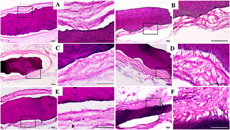
Figure 6.
HE staining of iTDM specimen harvested from the greater omentum after demineralization, embedding and section. In CLC groups (A: non-induced CLC; C: IFCM–induced CLC; E: MFCM-induced CLC) no evident attachment to the surface of iTDMs was formed, while periodontal ligament-like fibers were found to attach to the surface of iTDM with an angle in HERS groups. (B) Non-induced HERS cells formed the least amount of fibrous attachment to iTDM, IFCM-induced HERS cells (D) formed more, and MFCM-induced group (F) formed the most. The right column was the magnification of the black box in the left column, respectively. Scale bars: 100 μm.

