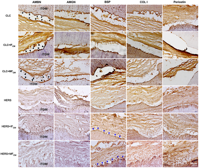
Figure 7.
Immunohistochemistry staining of AMBN, AMGN, BSP and COL I and Periostin. AMBN, AMGN, BSP and COL I were positively stained at the interfacial layers of iTDM and the tissues opposite to iTDM in CLC groups (indicated by black arrows). These indicated that enamel-like minerals may deposit on surfaces of iTDM seeded with CLC cells. Conversely, HERS cell groups showed negative expression of AMBN and AMGN but positive for BSP, COL I and Periostin. A thin layer at the surface of iTDM in HERS cell groups (indicated by blue arrows) was positively stained for BSP, COL I and Periostin. The fibers attached to the surface layer of the iTDM were also positive for COL I and Periostin. These indicated that cementum and periodontal ligament-like tissues were formed especially in IFCM and MFCM-induced HERS cells. Scale bars: 50 μm.

