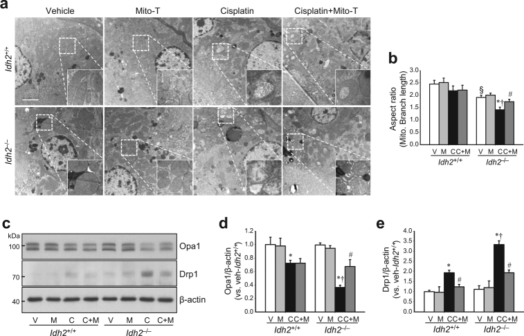
Fig. 5. IDH2 deficiency augments mitochondrial damage after cisplatin administration.
Idh2‒/‒ mice and wild-type (Idh2+/+) littermates were intraperitoneally injected with either cisplatin (C, 20 mg/kg B.W.) or 0.9% saline (vehicle, V) once. Some mice were treated with Mito-T (M, 0.7 mg/kg B.W.) daily, beginning 7 days before cisplatin injection and continuing until experiments were completed. a Two days after cisplatin administration, mitochondrial structures were examined by transmission electron microscopy (TEM). Higher magnification is shown by the dash-lined rectangles. Scale bar indicates 2 μm. b The mitochondrial aspect ratio [(major axis)/(minor axis)] was computed using 30 mitochondria per cell. c Expressions of OPa1 and Drp1 were determined by western blot analysis. β-actin was used as a loading control. d, e OPa1 (d) and Drp1 (e) band densities were measured using the ImageJ program. Results are expressed as means ± SE (n = 3–4 per group). Scale bars: a 2 µm. *p < 0.05 vs. respective V; #p < 0.05 vs. respective C; §p < 0.05 vs. V in Idh2+/+; †p < 0.05 vs. C in Idh2+/+

