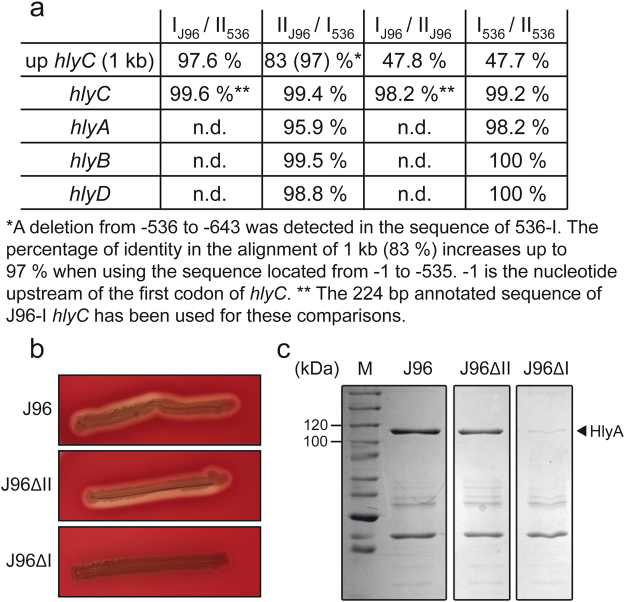
Figure 1.
Differential expression of the two hlyCABD operons present in the J96 strain. (a) Percentage of identity at the level of the nucleotide sequence between the operon hlyI and hlyII of J96 and 536. n.d.: not determined. (b) Hemolytic phenotype of J96, JFV16 (J96ΔII) and JFV21 (J96ΔI) strains on Columbia Blood agar plates. (c) Coomassie blue stained SDS-PAGE (10%) of secreted protein extracts from cultures grown in LB0 at 37 °C up to late-log phase (OD600 nm of 1.0) of the indicated strains. Lane M: molecular mass markers (size in kDa indicated along the left side). The band corresponding to α-hemolysin (HlyA) is indicated with an arrowhead. Full-length gel image is shown in Fig. S6.

