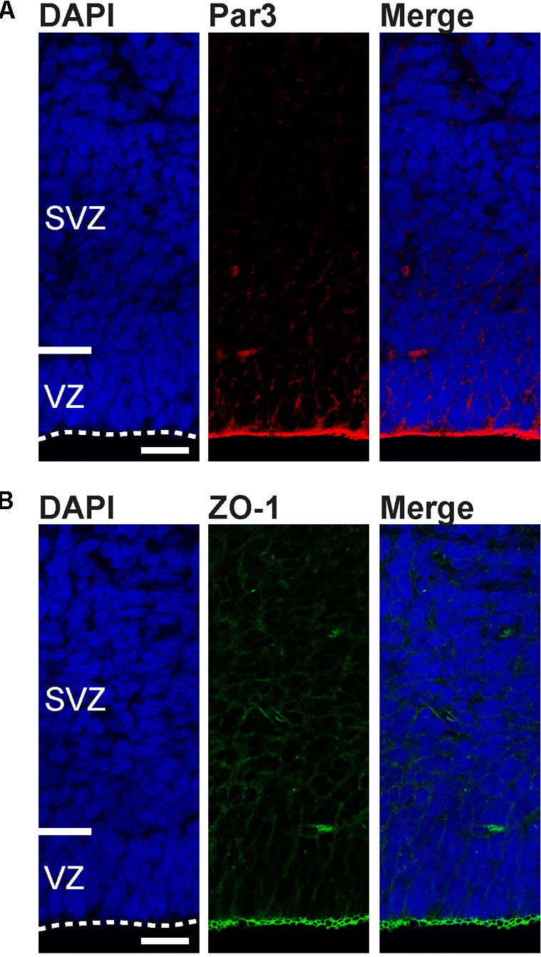
FIGURE 6.

Apical domain markers are concentrated at the developing tree shrew ventricular surface. (A,B) Immunofluorescence for Par3 (A, red), ZO-1 (B, green), and DAPI staining (blue) on 30 μm-cryosections of E37 tree shrew neocortex. Dashed lines indicate the ventricular surface. Scale bars, 50 μm. VZ, ventricular zone. SVZ, subventricular zone.
