Figure 5.

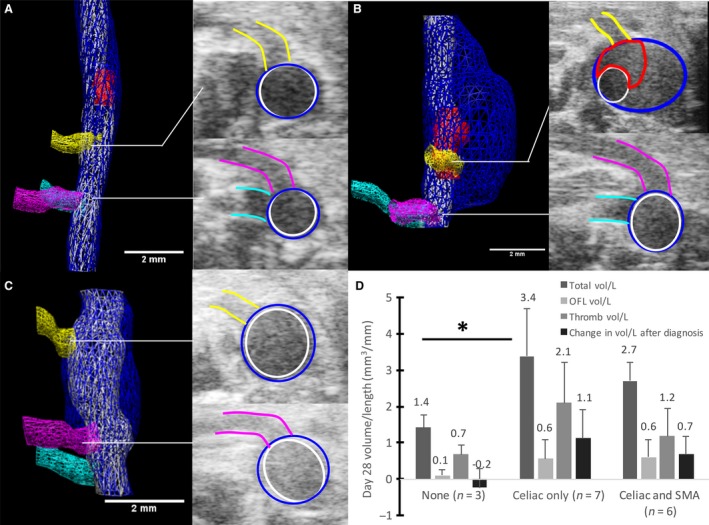
Effects of branching arteries on dissecting AAA growth. Volume segmentations of mice with no branching arteries (A), only the celiac artery branching into the expanded region of the vessel (B), and both the celiac artery and SMA branching into the expanded region (C). The celiac (yellow), SMA (pink), and right renal artery (cyan) are shown in all images. Open‐false lumens are segmented in red for (A) and (B), whereas the false lumen of (B) was entirely open and thus not segmented. Transverse US images show the celiac (top) and SMA (bottom) intersecting the aneurysm. Scale bars: 2 mm. When separated by branching arteries, total, open‐false lumen, and thrombus volume/length, animals with only the celiac artery have the largest volumes and greatest growth (D). The overall change in volume/length from day of diagnosis to day 28 was larger for the celiac only dissecting AAAs compared to the celiac and SMA group (*P ≤ 0.05). These plots represent the need to include branching vessel information when studying factors that influence dissecting AAA growth over time.
