
Figure 1.
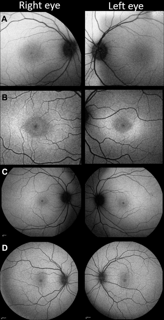
Fundus autofluorescence (FAF) changes in childhood-onset ABCA4-associated retinopathy. A, Patient 2 has a subtle increase in foveal FAF, while maintaining a small central zone of physiological hypoautofluorescence. B, In addition to the changes present in (A), a rhomboid zone of hyperautofluorescence is visible in patient 4. C, Peripheral macula imaging from patient 1 revealing a more extensive, diffuse hyperautofluorescent signal. D, A normal pattern of childhood autofluorescence shown for comparison.