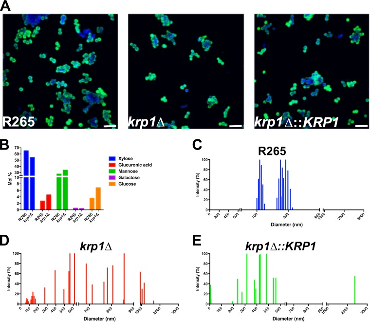
FIG 6 .
Morphological and structural proprieties of cell-associated and secreted polysaccharides from C. gattii strains. (A) Capsule transfer assay. C. neoformans cap67 cells were grown in YPD medium and incubated with secreted polysaccharides from WT and mutant C. gattii strains. For confocal microscopy, C. neoformans cells were labeled with Calcofluor white (blue) and MAb 18B7 (green). Bars, 20 µm. (B) Cell-associated polysaccharides were isolated from WT and krp1Δ cells, and their elemental composition was determined by GC-MS. (C to E) Cell-associated polysaccharide molecular dimension determination of WT, krp1Δ, and krp1Δ::KRP1 strains using DLS analysis. The cells were cultured in minimal medium for 72 h, and the cell-associated polysaccharides were extracted using DMSO. Each graph displays the range of polysaccharide fiber sizes representative of three independent analyses.

