The usage of the term “Sanderson's Polster” is cited in literature mainly in the first half of the 20th century. Sanderson-Damberg (1911) used the term Sanderson's polster, and probably Aschoff (1925) called similar structures as “Proliferationsknospe.”[1,2] “ Polster” is a German word that means a “cushion,” “pillow” and “bolster,” hence a metonymic occupational name for a maker or seller of pillows or a nickname for a plump man, and thus this word was probably used to denote based on the appearance of small secondary follicles within an enlarged follicle resembling a “cushion” or “pillow” attached to one end.
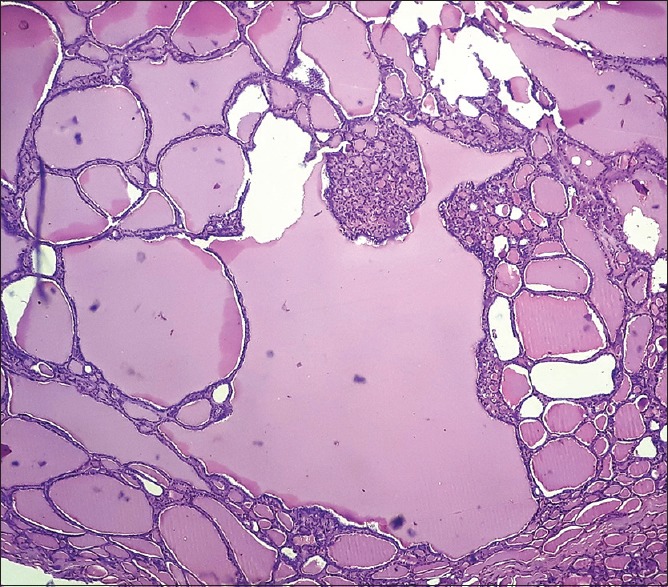
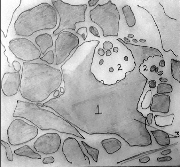
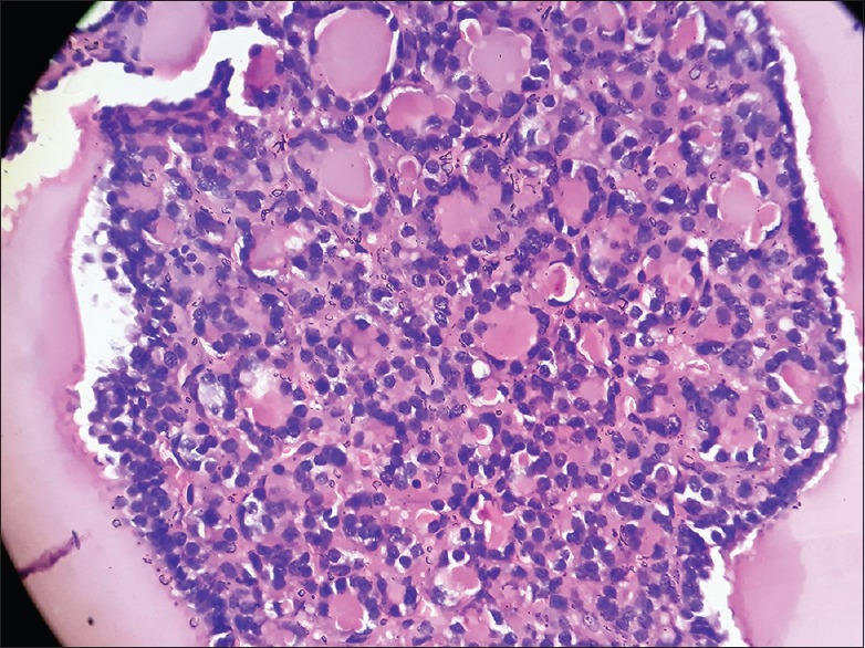
In normal thyroid gland or more commonly in hyperplastic conditions of thyroid gland such as nodular/multinodular goiter, adenomatoid goiter, or adenomatous hyperplasia, microscopically dilated thyroid follicles lined by cuboidal cells and filled with colloid may be seen. When colloid secretion increases in the small secondary follicles, their mass bulges into the follicular cavity and gives rise to a “Sanderson Polster” seen at one pole along the follicular wall of enlarged and dilated follicles.[3] These dilated follicles are usually 2–3 times larger than the surrounding follicles [Figure 1]. Figure 2 shows hand-drawn pictures corresponding to Figure 1. Sanderson's polster is composed mainly of small follicles lined by cuboidal cells [Figure 3]. Goormaghtigh and Thomas suggested the term “papilla” for these structures.[3] These secondary acini are nearly always lined with large epithelial cells, and the nuclei are conspicuous by their size. In an early study, Sugiyama et al. could not find “Sanderson's Polster” or “Proliferationsknospe” in the normal development of thyroid gland.[4]
Figure 1.

Photomicrograph of H&E section showing Sanderson polsters within an enlarged follicle (×40)
Figure 2.

Corresponding hand-drawn illustration of Figure 1: (1) The enlarged follicle, (2) secondary follicles (Sanderson polsters), and (3) colloid
Figure 3.

Photomicrograph of H&E section showing Sanderson polsters (×400)
Based on histochemical staining in aged murine thyroid tissue, Lee et al. opined that Sanderson's polsters, small follicles and irregularly-shaped follicles, were hyperactive as compared with markedly enlarged follicles.[5] Abundant colloid in large follicles was stained purple-red or dark blue-purple by PAS, while cellular areas composed of small follicles were pale-red or negative. In another study, Aeschimann et al. considered similar budding intraluminal protrusions as actively replicating, while flat neighboring patches were apparently resting. The results were drawn on the basis of p21 ras immunohistochemistry. p21 ras can be used as a growth marker. The areas of active growth show a significant increase in cellular p2l ras as compared to resting cells which contain very little p21 ras.[6]
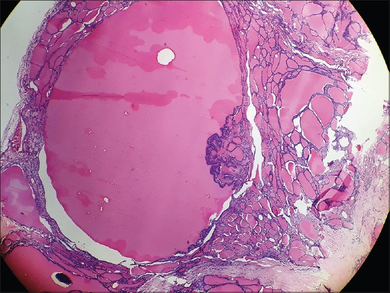
Instead of microfollicles at one pole, it is not unusual to find benign papillary formations protruding into the lumen of large follicles [Figure 4]. Such papillary protrusions should not be confused with papillary carcinoma of thyroid (PTC), as they lack the classical PTC nuclear features such as clearing of chromatin, nuclear enlargement, intranuclear cytoplasmic pseudoinclusions, nuclear grooving and nuclear overlapping.
Figure 4.

Photomicrograph showing multiple papillary fronds at one pole of large colloid filled thyroid follicle in a case of nodular colloid goiter
Despite the fact that these structures are sometimes described as areas of active growth, clinically majority remains entirely benign. However, familiarization with such terms may avoid an overdiagnosis of thyroid malignancy.
Financial support and sponsorship
Nil.
Conflicts of interest
There are no conflicts of interest.
Acknowledgment
Dr Sangeetha K Nayanar, Professor and HOD, Department of Clinical Laboratory Services and Translational Research, Malabar Cancer Center, Thalassery, Kerala.
REFERENCES
- 1.Sanderson-Damberg E. The thyroid glands from 15-25. Year of life from the North German plain and coastal area as well as from Bern. Frankfurt Z Path. 1911;6:312–34. [Google Scholar]
- 2.Aschoff L. Jena: Gustav Fischer; 1925. Lectures on Pathology Held at the Universities and Academies of Japan in 1924; pp. 1–309. [Google Scholar]
- 3.Goormaghtigh N, Thomas F. The functional reactions of the human thyroid: A Contribution to its histophysiology. Am J Pathol. 1934;10:713–30. [PMC free article] [PubMed] [Google Scholar]
- 4.Sugiyama S, Taki A, Nakano A, Sugiyama N, Yamamoto Y. Histological studies of the human thyroid gland in middle and late prenatal life. Okajimas Folia Anat Jpn. 1959;33:75–84. [Google Scholar]
- 5.Lee J, Yi S, Kang YE, Kim HW, Joung KH, Sul HJ, et al. Morphological and functional changes in the thyroid follicles of the aged murine and humans. J Pathol Transl Med. 2016;50:426–35. doi: 10.4132/jptm.2016.07.19. [DOI] [PMC free article] [PubMed] [Google Scholar]
- 6.Aeschimann S, Kopp PA, Kimura ET, Zbaeren J, Tobler A, Fey MF, et al. Morphological and functional polymorphism within clonal thyroid nodules. J Clin Endocrinol Metab. 1993;77:846–51. doi: 10.1210/jcem.77.3.8370709. [DOI] [PubMed] [Google Scholar]


