
Fig. 3. NRGs induce in vitro resistance to PI3K inhibition in HER2-amplified and/or PIK3CA-mutant BC cells, and this is dependent on HER3 activation.
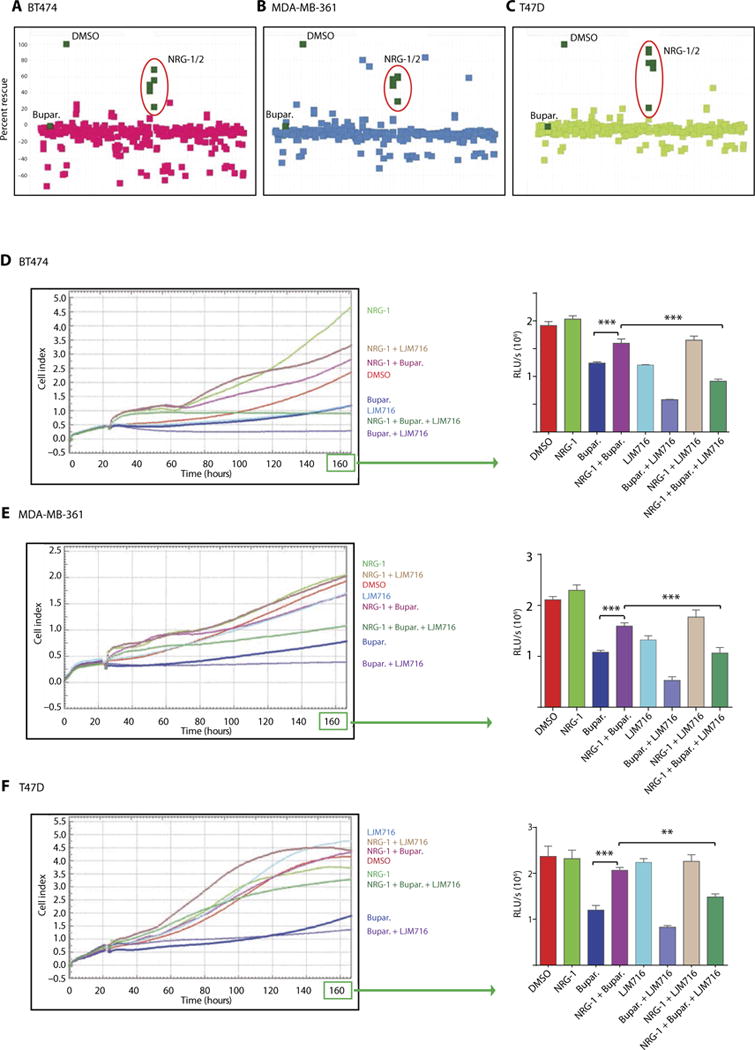
(A to C) A library of secreted proteins was screened for their ability to rescue the inhibition of BT474 (A), MDA-MB-361 (B), and T47D (C) BC cell growth in vitro by buparlisib. Each dot represents the average of three replicates of cells conditioned with the same supernatant. “DMSO” represents cells treated with dimethyl sulfoxide control in the absence of secreted proteins (rescue, buparlisib-treated cells in the presence of growth factor relative to control). (D to F) In vitro xCELLigence growth assay of BT474 (D), MDA-MB-361 (E), or T47D (F) BC cells exposed under the indicated conditions for the indicated time. The y axis is cell index defined as the relative change in measured electrical impedance. The graphs on the right show CellTiter-Glo luminescence viability measurements at the end of the experiments [one-way analysis of variance (ANOVA)/Tukey test, **P = 0.0009, ***P < 0.0001]. Data are means ± SD.