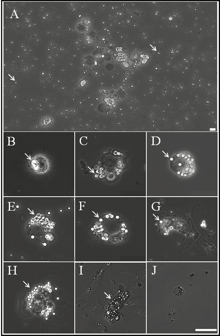
Fig. 4.
Light micrographs showing blood cells cultured with carboxylate-modified polystyrene latex beads. (A) Overall shape of cultured hemocytes and beads (indicated by white arrows). (B–J) Cultured granulocytes at 6 h, 9 h, 12 h, 18 h, 1 day, 2 days, 7 days, 10 days, and 14 days. Scale bar = 10 µm. By 7 d of culture, most granulocytes showed polymorphic glittering vacuoles (beads), which were closely packed within the cytoplasm (indicated by white arrows).

