Figure 1.
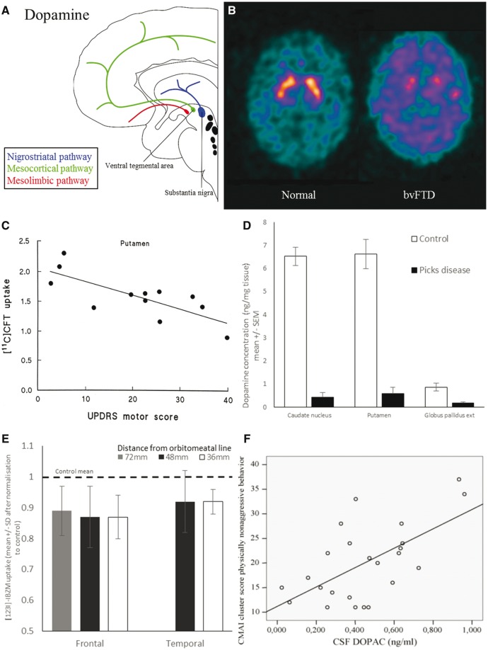
Dopamine deficits in FTD. (A) Schematic illustration of dopaminergic pathways. (B) Ioflupane SPECT scan showing loss of pre-synaptic dopaminergic neurons in the striatum of FTD compared with normal scan. (C) Loss of dopaminergic neurons in the putamen (measured by 11C-CFT-PET) correlates with severity of extra-pyramidal motor symptoms (Unified Parkinson’s Disease Rating Scale motor score). From Rinne et al. (2002). Reprinted with permission from Wolter Kluwer. (D) Dopamine levels are reduced in the caudate, putamen and globus pallidus. Graph of data from Kanazawa et al. (1988). Reprinted with permission from Elsevier. (E) There is loss of D2 dopamine receptors in the frontal lobes (as measured by 123I-IBZM-PET). Graph of data from Frisoni et al. (1994). Reprinted with permission from Elsevier. (F) CSF DOPAC levels (3,4-dihydroxyphenylacetic acid, a dopamine metabolite) correlate with behavioural disturbance. From Engelborghs et al. (2008). Reprinted with permission from Elsevier.

