
Figure 1.
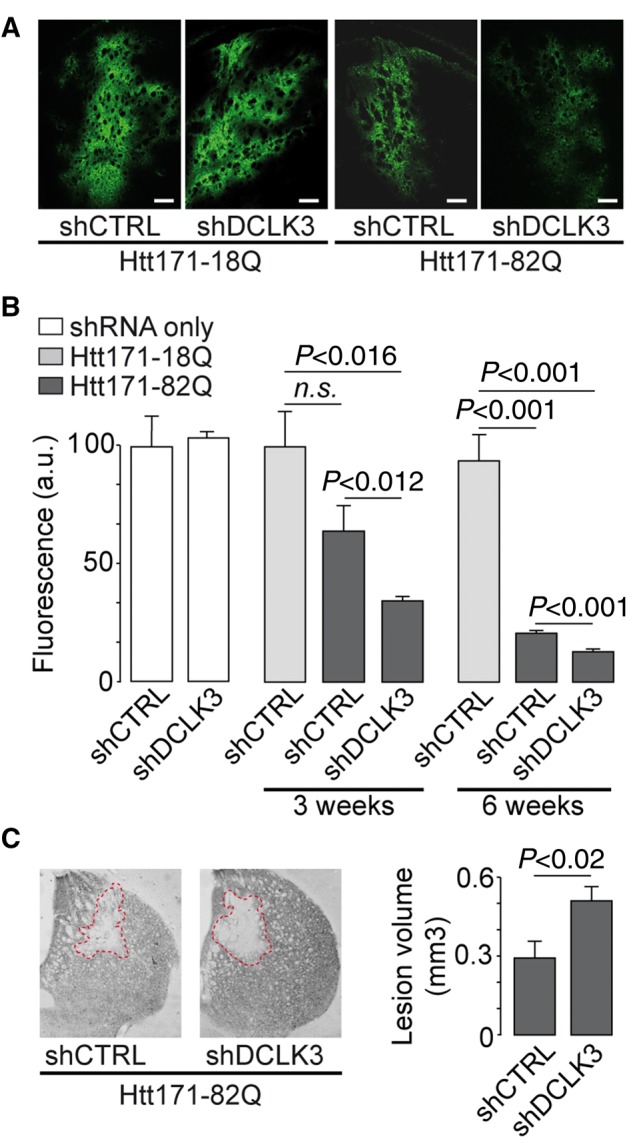
DCLK3 knockdown increases the toxicity of mHtt in vivo. Mice received lentiviral vectors encoding a shRNA targeting either DCLK3 (shDCLK3) or luciferase (shCTRL, the control). The shRNA-encoding constructs also contained the coding sequence for GFP. (A) Representative images of GFP expression in striatal sections from mice infected with LV-Htt171-82Q or LV-Htt171-18Q with LV-shDCLK3 or its control (LV-shCTRL) (3 weeks post-infection). (B) Quantification of fluorescence as an index of neuronal integrity in mice infected with LV-Htt171-18Q mixed with LV-shCTRL (control levels of GFP) or LV-Htt171-82Q mixed with LV-shDCLK3 or LV-shCTRL. Quantification was performed 3 and 6 weeks after infection. The Htt171-82Q-induced loss of GFP was exacerbated by LV-shDCLK3. (C) Brains were processed for histological evaluation 6 weeks after infection, with DARPP32 histochemistry used to detect Htt171-82Q toxicity. Left: Typical coronal sections of mouse brain, with areas of staining depletion observed in the presence of Htt171-82Q. Right: Quantitative determination of the size of the striatal lesions in the two groups. Infection with shDCLK3 significantly leads to increased lesion volume, as shown with DARPP32. Results are expressed as means (n = 7–10/group) ± SEM. Mann-Whitney and Kruskall-Wallis tests in B and paired Student’s t-test in C. Scale bars in A = 0.25 mm; B = 0.5 mm.