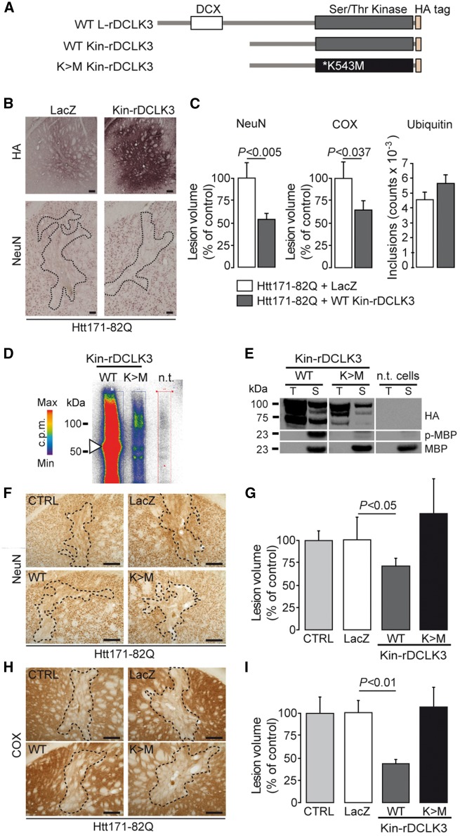
Figure 4.
The active kinase domain of rDCLK3 is sufficient to protect against mHtt in the striatum in vivo. (A) Schematic illustration of DCLK3 and Kin-rDCLK3-HA constructs with and without the K543M mutation. (B) Mice received bilateral injection of LV-Htt171-82Q mixed with either LV-LacZ (control) or a lentivirus encoding the kinase domain of DCLK3 (LV-Kin-rDCLK3). Six weeks after infection, brains were processed for histological evaluation (B) of NeuN, HA and COX (not shown) levels. HA staining (B) shows expression of Kin-rDCLK3-HA in the striatum. (C) Quantification of the volumes of the Htt171-82Q-induced striatal lesions by staining for NeuN, COX and ubiquitin. (D) Autophosphorylation experiments with ATP [γ-32P] after the co-immunoprecipitation of wild type or mutant Kin-rDCLK3 with anti-HA antibody followed by SDS-PAGE, showing the incorporation of radioactive 32P into the protein. Colours indicate the radioactivity levels from white (no signal) to red (highest). (E) Assays of kinase activity, with MBP used as a kinase substrate after the of Kin-rDCLK3-HA, wild-type (WT) and mutant K543M forms (K > M) and detection of phosphorylated MBP (p-MBP) by western blotting. T = input (total protein extract before immunoprecipitation), S = sample test; n.t. = non-transfected cells (control). (F and H) Photomicrographs showing striatal lesions in mice injected with LV-Htt171-82Q mixed with LV-LacZ (control, CTRL), LV-Kin-rDCLK3 (WT) or lentivirus encoding the mutant Kin-DCLK3 form with the K543M (K>M) substitution. (G and I) Histograms representing the quantification of the volumes of Htt171-82Q-induced lesions using NeuN (G) and COX (I) staining. Scale bars = 0.2 mm. Results are expressed as means (n = 7–11/group) ± SEM. P-values indicate the level of significance (unpaired Student’s t-test, control versus wild-type).

