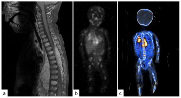
Figure 2. Imaging in Neuroblastoma.
Imaging in neuroblastoma must be multimodal to accurately locate and characterize the primary tumor with cross-sectional imaging and locate metastatic disease with MIBG. A) An MRI showing a paraspinal mass invading the spinal canal across many thoracic levels, causing spinal cord compression. Also note the metastatic involvement of multiple vertebral bodies. B) 123I-metaiodobenzylguanidine (MIBG) scan showing the primary thoracic tumor and revealing wide spread metastatic disease involving the bones. C) Single-photon emission computed tomography (SPECT) combining MIBG and CT to better localize the MIBG uptake.

