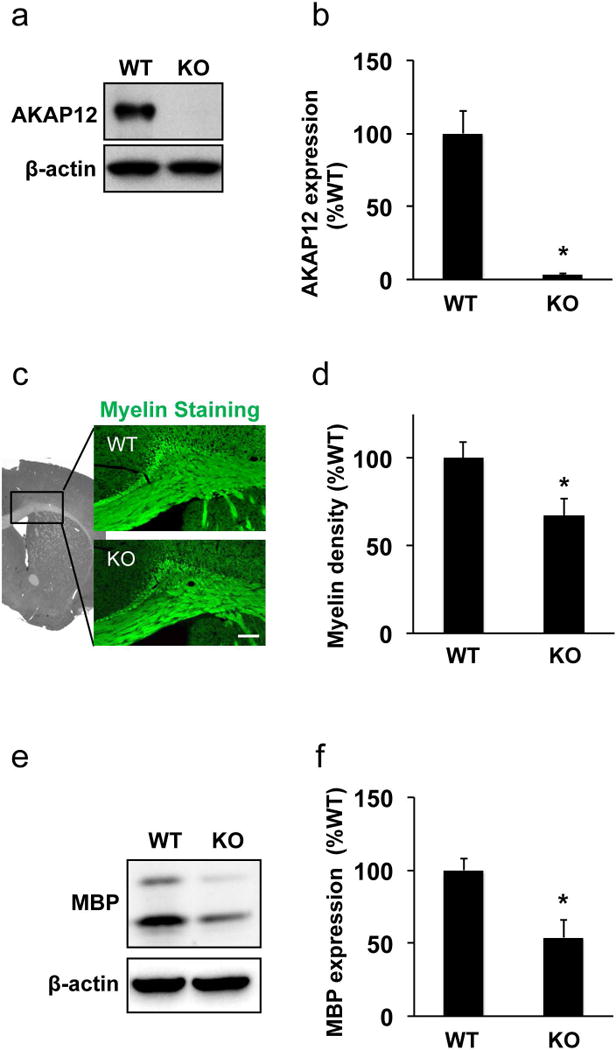
Figure 1. Loss of white matter myelin in AKAP12 knockout mouse.

(a-b) Western blot using anti-AKAP12 antibody confirmed AKAP12 expression only in wild-type mouse cerebral white matter (i.e. corpus callosum region). β-actin was used as an internal control. Values are mean ± SD. N=6. *P<0.05. (c-d) Myelin staining using the FluoroMyelin kit showed white matter lesion with myelin damage in AKAP12 knockout (KO) mice. The densitometric analysis confirmed that myelin density in wild-type mice (WT) was significantly higher compared to AKAP12 KO mice. Bar=100 μm. Values are mean ± SD. N=5. *P<0.05. (e-f) White matter samples were subjected to western blot analysis. Expression of myelin-basic protein (MBP), a major protein in white matter myelin, was significantly larger in WT mice. β-actin was used as an internal control. Values are mean ± SD. N=5. *P<0.05.
