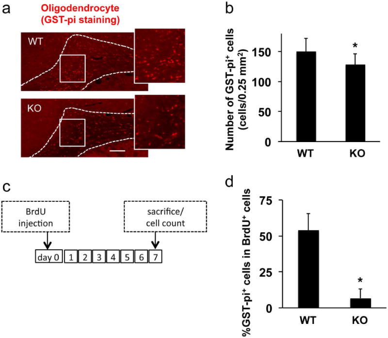
Figure 3. Oligodendrocyte loss in AKAP12 knockout mouse.

(a-b) Immunostaining using anti-GST-pi antibody showed that the number of oligodendrocytes in white matter (corpus callosum area) in AKAP12 knockout (KO) mice was significantly lower compared to wild-type (WT) mice. Bar=25 μm. Values are mean ± SD. N=5. *P<0.05. (c) Protocols for in vivo OPC-to-oligodendrocyte differentiation assay. Seven days after BrdU injection, brains were taken out and subjected to immunostaining. (d) Double staining of BrdU with anti-GST-pi antibody (oligodendrocyte marker) showed that AKAP12 KO mice exhibited low level of newly generated oligodendrocytes. Values are mean ± SD. N=5. *P<0.05. Please see Supplementary Figure S4 for representative images for GST-pi/BrdU double-staining.
