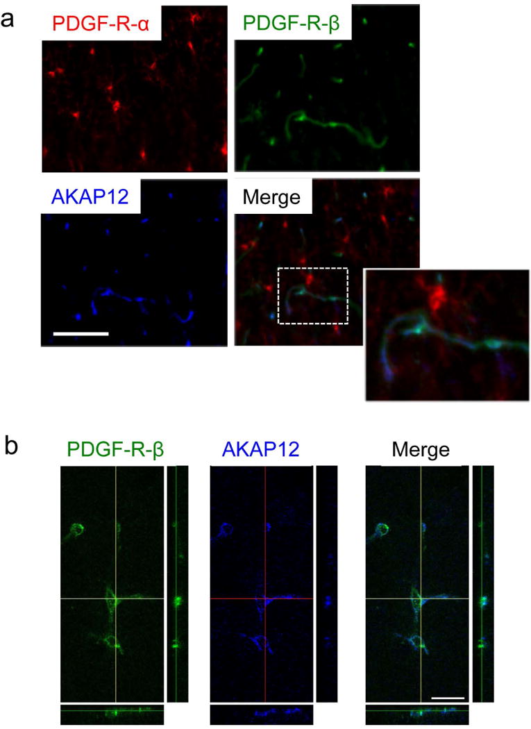
Figure 4. AKAP12 expression in pericytes in vivo.

(a) Immunostaining showed that AKAP12 was not expressed in OPCs (PDGF-R-α positive cells) in the corpus callosum region in wild-type mouse brain sections. Instead, AKAP12 was confirmed to be expressed in pericytes (PDGF-R-β positive cells) in the corpus callosum region in wild-type mouse brain sections. Importantly, AKAP12-expressing pericytes were closely located to OPCs in the corpus callosum region. Bar=100 μm. (b) Double-staining of PDGF-R-β and AKAP12 using confocal microscopy confirmed that pericytes express AKAP12. Bar=20 μm. Please see Suppl Figure S6 for additional data from immunostaining with another pericyte marker CD13.
