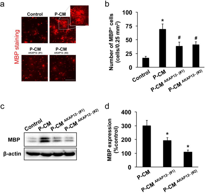
Figure 7. Pericytic AKAP12 for OPC differentiation in vitro.

Cultured OPCs were maintained in control media, pericyte-conditioned media (P-CM), or conditioned media from AKAP12-deficient pericytes (P−CMAKAP12-). Five days later, those OPCs were used for immunostaining or western blotting analyses. (a-b) Immunostaining with anti-MBP (oligodendrocyte marker) antibody showed that P-CM increased the number of MBP−positive cells, but P-CMAKAP12- showed less effect on OPC maturation. Bar=100 μm. Values are mean ± SD. N=5. *P<0.05 vs control, and #P<0.05 vs P-CM. (c-d) The levels of MBP expression in P-CM-treated OPCs were also lower compared to ones in P−CMAKAP12- -treated OPCs. Values are mean ± SD. N=4. *P<0.05 vs P-CM.
