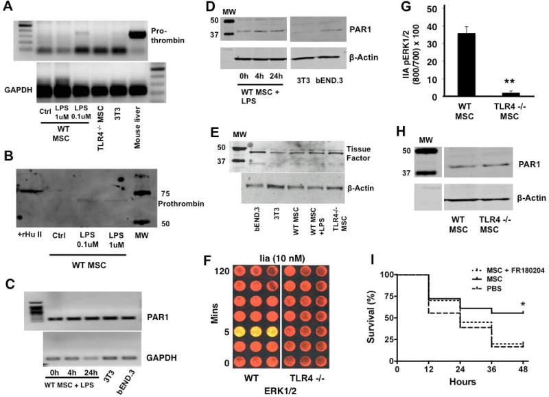
Figure 3. TLR4 stimulation induces prothrombin secretion by MSCs and regulates signaling through PAR1.
WT MSCs demonstrated evidence of prothrombin gene expression upon stimulation with 100 nM of LPS, which was not seen in TLR4 −/− MSC or 3T3 fibroblasts. Mouse liver cDNA was used as a positive control for prothrombin (A). Conditioned media from MSCs stimulated with LPS demonstrated the presence of prothrombin protein as detected by Western blotting. Recombinant human prothrombin was used as the positive control for these studies (B). Reverse transcriptase-PCR demonstrated that MSCs expressed the transcript for PAR1 (C), and Western blotting confirmed that MSCs expressed the PAR1 protein (D). There was no appreciable change in PAR1 expression by MSCs with LPS stimulation (3T3, mouse embryonic fibroblast cell line; bEND.3, mouse brain endothelial cells line used as positive control). MSCs also were shown to express tissue factor by Western blotting (E, bEND.3 cell line used as positive control). Thrombin stimulation (10 nM) led to significant ERK1/2 phosphorylation at 5 min in WT MSCs but not in TLR4 −/− MSCs (F, G, **p < 0.01 for TLR4 −/− MSCs vs WT MSCs; n = 3 per group per time point), despite both WT and TLR4 −/− MSC qualitatively expressing similar amounts of PAR1 protein by Western blotting (H). Using the in vivo model, we determined that inhibition of ERK1/2 with FR180204 eliminated the therapeutic efficacy of WT MSCs, suggesting that this central signaling pathway is required for MSC-derived protection (I, *p < 0.05 for MSC vs PBS treated group, n = 18 – 20 per group).

