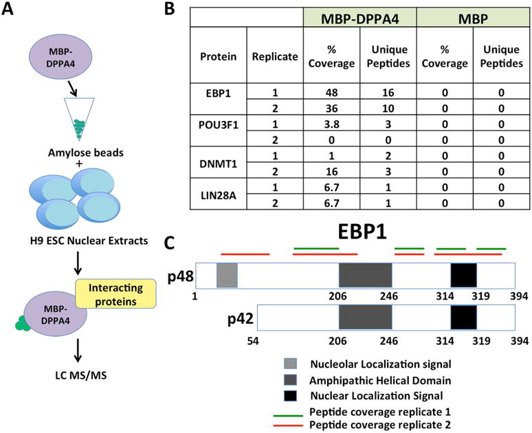
FIGURE 1. A DPPA4 proteomics screen identifies candidate interacting proteins, including EBP1.

(A) MBP or MBP-DPPA4 was bound to amylose beads and incubated with nuclear extracts from H9 ESCs. Unbound proteins were washed off and interacting proteins were identified through LC MS/MS. Two biological replicates were performed. (B) Table of representative proteins identified through LC-MS/MS to be bound to DPPA4-MBP at 0.05 FDR. (C) Mass spectrometry peptide coverage of EBP1 from two biological replicates shows coverage of both isoforms of EBP1. Both the unique region of p48, within the first 54 amino acids, and regions shared by p42 and p48 isoforms were detected.
