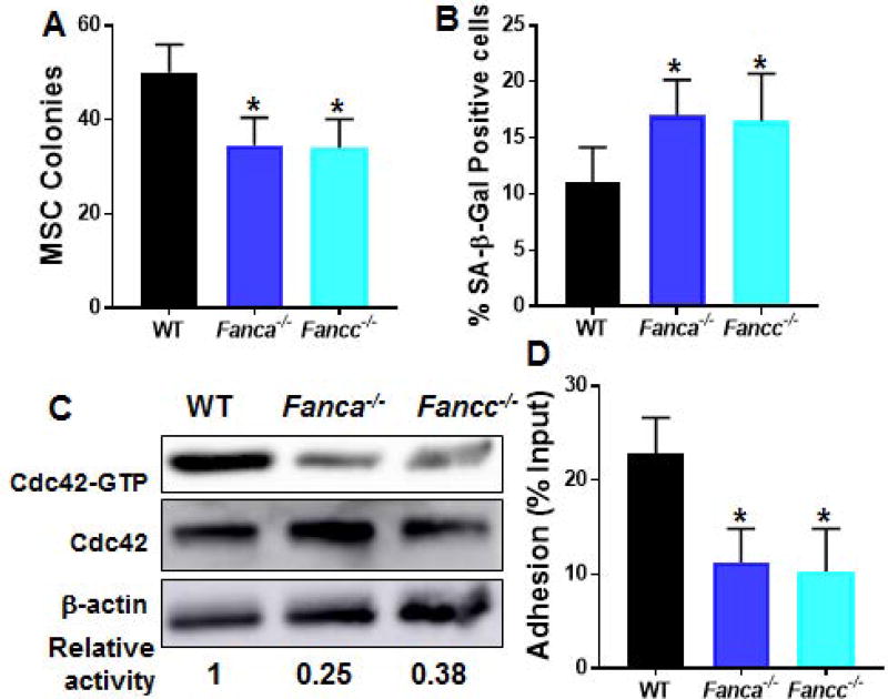
Fig. 1. Reduced Cdc42 activity and adhesion defect in Fanca−/− and Fancc−/− MSCs.
(A) Fanca−/− and Fancc−/− MSCs exhibit defective proliferation in vitro. MSCs isolated from wild-type (WT), Fanca−/− or Fancc−/− mice were cultured in MSC medium followed by MSC colony forming efficiency (CFE) assay. Numbers of colonies formed were enumerated on day 12 in triplicate from five individual WT, Fanca−/− or Fancc−/− mice. Quantification are shown. (B) Fanca−/− and Fancc−/− MSCs are prone to senescence. Cells described in (A) were passaged for 3 times in vitro followed by SA-β-gal staining. Percentages of the cells stained positive for SA-β-gal were quantified by counting a total of 100 cells in random fields per well. (C) Decreased Cdc42 activity in Fanca−/− and Fancc−/− MSCs. MSCs isolated from WT, Fanca−/− or Fancc−/− mice were cultured in MSC medium and passaged for 3 times. Whole cell lysates (WCL) was then extracted from the cells and subjected to Western blotting using antibodies against Cdc42. The level of active, GTP-bound Cdc42, total Cdc42 and β-actin were determined. The relative levels of active Cdc42 are indicated below the blot. (D) WT HSPCs co-cultured on Fanca−/− or Fancc−/− MSCs display decreased adhesion. Sorted LSK (Lin−Sca1+c-kit+) cells from WT mice were added onto confluent WT, Fanca−/− or Fancc−/− BM-derived MSCs and incubated for 4 hours at 37°C; non-adherent cells were then subjected to colony forming cell (CFC) assay. Numbers are given as the percentage of input CFCs with three independent experiments in triplicates in each experiments (n=5). *p<0.05.

