
Figure 2. MRI data showed comparable functional improvements of cardiac functions of rats injected with ESC-CMs and iPSC-CMs.
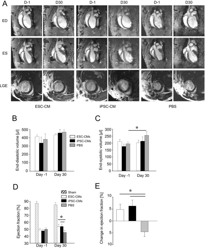
Quantitative MRI was used to assess left-ventricular ejection fractions of ischemic rats receiving ESC-CMs (n = 8), iPSC-CMs (n = 8), or PBS (n = 7). (A) The top row shows representative 2-chamber long-axis views at end-diastole (ED) one day prior to (D-1) and 30 days after (D30) cell implantation or PBS. The middle row shows the same hearts at end-systole (ES), whereas the bottom row shows corresponding late gadolinium enhancement (LGE) images at mid-ventricular level. (B) End-diastolic volumes increased in all groups but were not significantly different. (C) End-systolic volumes increased significantly in the PBS group (mean ± SEM; P=0.03), but the increases were not significant in ESC-CM and iPSC-CM groups. (D, E) Ejection fractions increased in ESC-CM and iPSC-CM groups but declined in the PBS group (mean ± SEM; P=0.03).