
Figure 6.
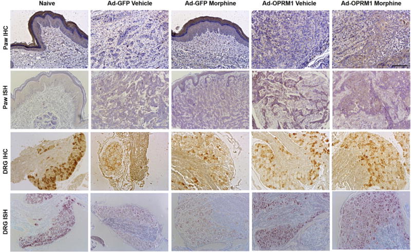
Ad-OPRM1 treatment rescued mu-opioid receptor expression in both paw cancer and corresponding DRG. Representative sections of immunohistochemical (IHC) staining and in situ hybridization (ISH) from paw cancer and DRG (L4–L5) for each treatment group are shown. The treatment group is indicated at the top of each column. Rows 1 and 2 show IHC and ISH stains of the paw cancer, which represent the expression of mu-opioid receptor protein and Oprm1 transcript, respectively. The full-thickness paw section is shown in the naive group and Ad-GFP morphine group. The dermal layer typically expresses mu-opioid receptor and is shown in this figure with positive brown staining. The cancer was inoculated directly below the dermal layer, as shown in the sections from the Ad-GFP morphine group. Brown staining within the cancer represents positive mu-opioid receptor protein expression (row 1) and Oprm1 transcript expression (row 2). Rows 3 and 4 show IHC and ISH stains from the DRG. DRG sections from the Ad-GFP vehicle and Ad-GFP morphine group have muted expression of mu-opioid receptor protein compared with the naive, Ad-OPRM1 vehicle, and Ad-OPRM1 morphine group (row 3, positive expression is represented by brown stain). Transcript expression is also muted in the Ad-GFP vehicle and Ad-GFP morphine groups compared with the other 3 groups (row 4, positive expression is represented by purple stain). Black bar = 100 μm.