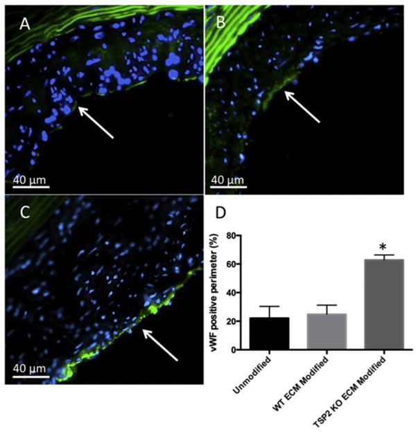
Fig. 4. TSP2 KO ECM modified grafts have increased endothelialization after 4 weeks in vivo.
Detection of vWF (green color) was performed after 4 weeks implantation on mid-graft sections of unmodified (A), WT ECM modified (B), and TSP2 KO ECM modified (C) grafts to visualize endothelial cells (Zeiss). (D) Quantification by Image J showed increased vWF-positive percentage of the lumen perimeter on TSP2 KO ECM modified grafts. n = 4, *p < 0.05. Arrows indicate luminal surface. (For interpretation of the references to colour in this figure legend, the reader is referred to the web version of this article.)

