Fig. 2.
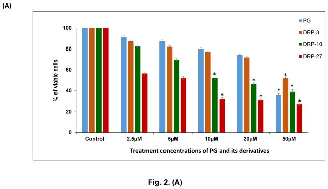
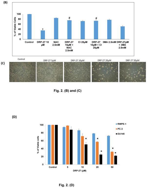
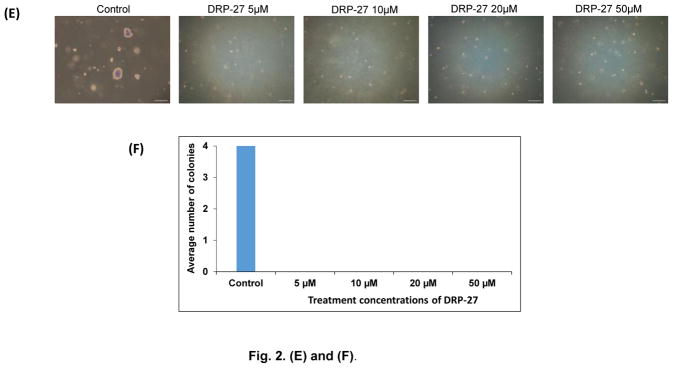
Cell viability assay of PG and its derivatives on prostate cancer cells: (A) Derivatives of PG, especially DRP-27, inhibit the proliferation of prostate cancer cells (LNCaP) more effectively than PG as determined by MTT assay. Data shown as mean ± SD. *P<0.05 compared to control group. (B) Anti-proliferative activity of DRP-27 against LNCaP cells were abrogated by NAC and CI. *P<0.001 compared to control. #P<0.01 compared to DRP-27 treatment group. (C) Morphological changes in LNCaP cells after treatment with DRP-27. Cells were treated for 48 h with various concentrations of DRP-27. Morphological changes were observed using microscopy (×20 magnification). (D) Cytotoxicity evaluation of DRP-27 on normal prostate cells (RWPE-1) and androgen-independent prostate cancer cells (PC-3 and DU145). Results showed that DRP-27 has minimal toxic effects on RWPE-1 compared to PC-3 and DU145 PCa cells. *P<0.05 compared to RWPE-1 cells. DRP-27 treatment reduces anchorage-independent growth of LNCaP cells: (E) Soft agar assay results show that DRP-27 significantly inhibited the colony formation ability of LNCaP cells. Colonies were detected using crystal violet staining. (F) Quantitative analysis of clonogenesis in LNCaP cells against different concentrations of DRP-27. Data are shown as mean ± S.D.


