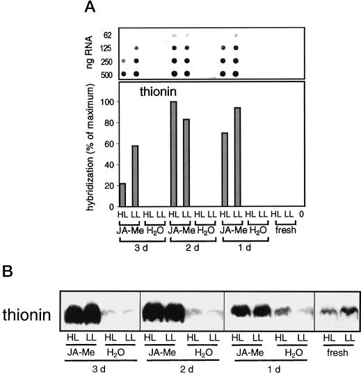
Figure 6.
Leaf thionin: mRNA and protein accumulation. A, Dot-blot analysis of poly(A)-rich mRNA. Application of JA-Me, light treatment, and analysis of poly(A)-rich mRNA were performed as described in the legend of Figure 2A, except that hybridization was done with a homologous probe to leaf thionin. B, Western-blot analysis of total protein extracts. Total protein extracts (15 μg of protein per lane) were analyzed by western blot probed with anti-leaf thionin following SDS-PAGE (20% [w/v] gels). The figure is composed of two western blots that were performed in parallel and treated identically (see also legend to Fig. 4).

