Fig. 1.

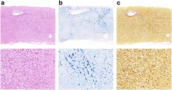
Liver biopsy histology of case 1. Histological sections stained with a H&E, b Perls, and c Van Gieson’s stain showing significant stainable iron, without pathology

Liver biopsy histology of case 1. Histological sections stained with a H&E, b Perls, and c Van Gieson’s stain showing significant stainable iron, without pathology