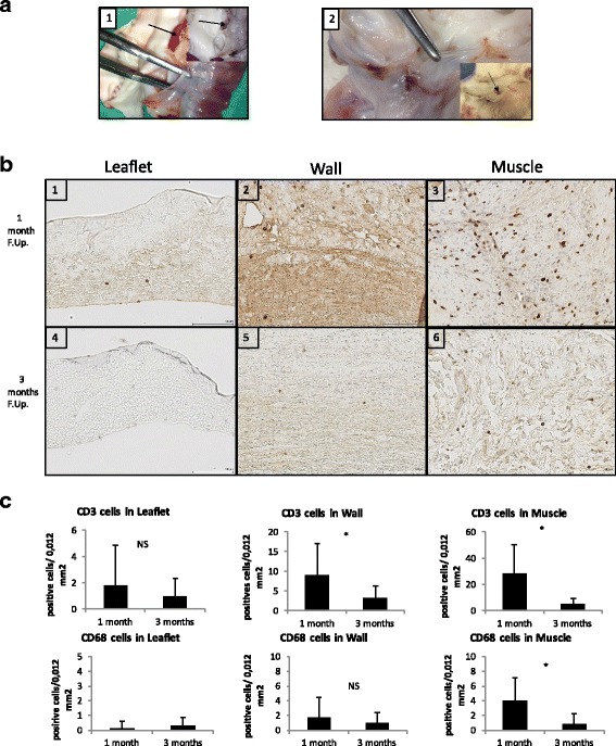
Fig. 5.

DPV in vivo results: macroscopic assessment and CD3/CD68 infiltration. a: Macroscopic views of explanted valves at one month (1) and three months (2) follow-up. The forceps indicate translucent and flexible leaflets. The black arrow in 1 shows thrombi formation and in 2, a micro calcification in the posterior sinus. b: Representative histological findings of CD3 staining of DPV at one month (first line) and three months (second line) of follow-up in leaflet (1;4), wall (2;5) and muscle (3;6). Note staining was the highest in muscle at one month (3) and reduction of staining at three months in comparison to one month (1- > 4 for leaflets/2- > 5 for wall and 3- > 6 in muscle). Scale bar: 100 μm. c: Results of histomorphometry for CD3 (first line) and CD68 (second line) staining in the three portions of DPV at one month and three months showing a decrease of inflammatory reaction at three months in all portions of the valve and significantly (*: p < 0,05) in wall and muscle portions for CD3 infiltration and in wall portion for CD68 infiltration. Low inflammation in the leaflet, the wall and muscle portions was found at 3 month: for CD3 infiltration: respectively 0,98 +/− 1,36; 3,1+/− 3,28 and 5,08 +/− 4,35 cells/0,12 mm2. and for CD68 infiltration: respectively CD68: 0,32 +/− 0,69; 0,93 +/− 1,44 and 0,87 +/− 1,34 cells/0,12mm2)
