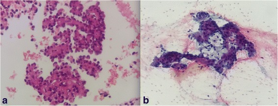
Fig. 3.

Histopathologic plates analysis of solid pseudopapillary tumor. a Cellular, single cells, small loose clusters, and scattered intact papillary structures with delicate fibrovascular cores, finely granular cytoplasm, and nuclei with fine chromatin. b Well-differentiated epithelial neoplasm, with papillary structures
