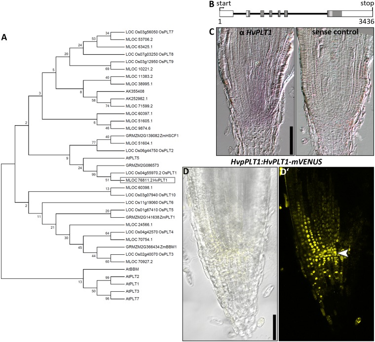
Fig 4. PLT phylogenetic tree, HvPLT1 gene structure, promoter activity and protein localization in the root meristem of the barley cv. Golden Promise 8 DAG.
(A) Phylogenetic tree of PLT homologue proteins. (B) Genomic structure of the HvPLT1 coding sequence; boxes represent exons, black horizontal lines represent introns; dark gray boxes indicate coding sequence for AP2 domains, light gray boxes indicate coding sequence for the linkers between AP2 domains. (C) Representative picture of RNA in situ hybridizations with a probe for HvPLT1 (purple staining, C)) or the respective sense probe (C’)). (D) Representative picture of the HvpPLT1:HvPLT1-mVENUS emission in the root meristem; transmitted light and mVENUS emission (D)), mVENUS emission only (D’)); arrow head in D’) points to the QC; hand sections; seven independent transgenic lines were examined and exhibited similar expression patterns; scale bars 100 μm.

