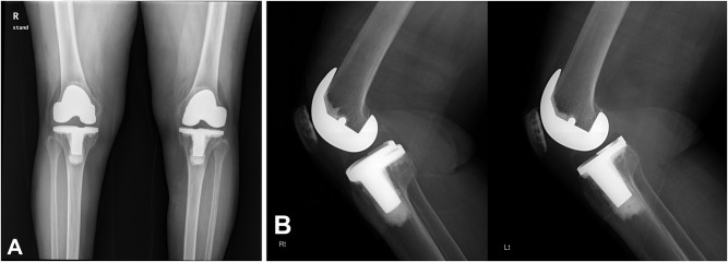
Fig. 3.
A-B Radiographs of both knees of a 53-year-old woman with end-stage osteoarthritis. (A) AP radiograph of both knees taken 15 years after surgery revealing the NexGen PS prosthesis performed with a computer-assisted technique (right knee, shown in the left image) and the NexGen PS prosthesis done with the conventional technique (left knee, shown in the right image) are embedded solidly in a satisfactory position. No radiolucent line or osteolysis is demonstrated adjacent to the tibial component in either knee. (B) Lateral radiographs of the same knees show the absence of radiolucent lines and osteolysis around the femoral, tibial, and patellar components in both knees. (The radiograph of the left knee has been flipped for the sake of better comparison.)

