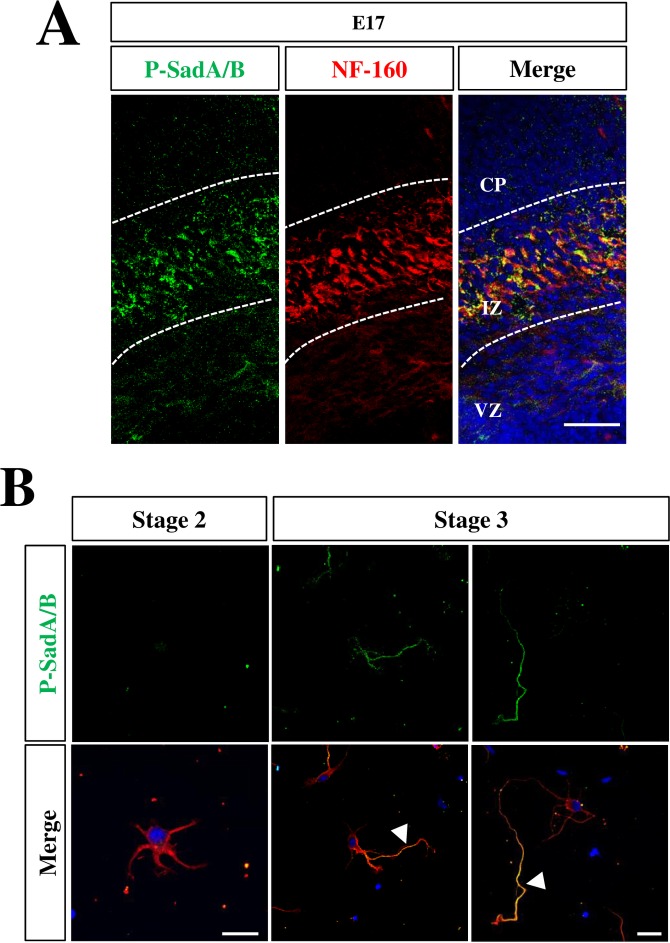
Fig 6. Active SadA and SadB are restricted to the axons of polarized neurons.
(A) Coronal sections from the brains of E17 mouse embryos were stained with Hoechst 33342 (blue) an antibody detecting active SadA and SadB phosphorylated at Thr175 and Thr187, respectively, (P-SadA/B, green) and the Tuj1 (red) antibody. (B) Cortical neurons from E18 rat embryos were analyzed at 1, 2 and 3 days in vitro (d.i.v., stage 2 to 3) by staining Hoechst 33342 (blue) an antibody detecting active SadA and SadB phosphorylated at Thr175 and Thr187, respectively, (P-SadA/B, green) and the Tuj1 (red) antibody. Early and late stage 3 neurons are shown. Axons are marked by arrowheads. The scale bars are 20 μm (A) and 50 μm, respectively (B).

