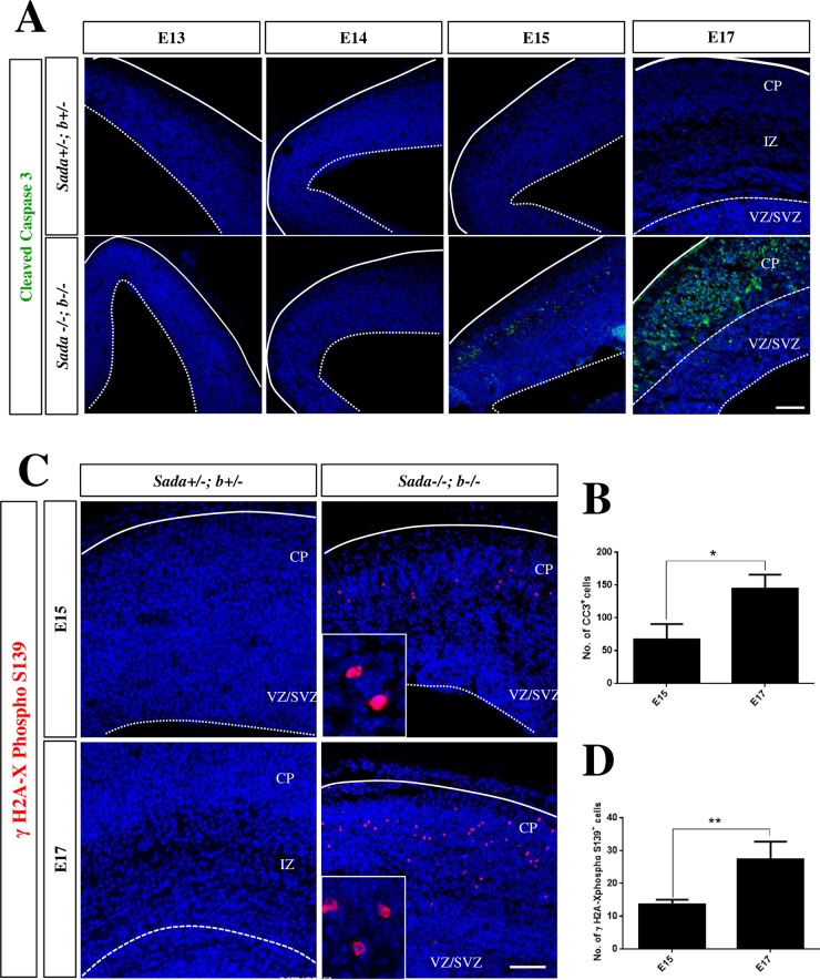
Fig 7. Increased apoptosis in the Sada-/-;Sadb-/- knockout brain.
(A, C) Coronal sections from the cortex of E13, E14, E15 or E17 embryos with the indicated genotypes were stained with an anti-cleaved caspase-3 (A, green) or anti-phospho-Ser139-γH2A.X antibody (B, red) and Hoechst 33342 (blue). (B, D) The number of nuclei per section positive for cleaved caspase-3 (B) or phospho-S139-γH2A.X (D) was determined in the cortex at the indicated stages. No signals for phospho-Ser139-γH2A.X were detectable in heterozygous controls. Values are means ± s.e.m., n = 3 different embryos, Student’s t-test n between E15 and E17 (*, p<0.05; **, p<0.01). The scale bars are 50 μm.

