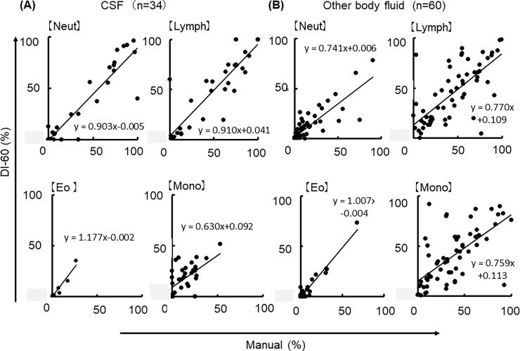
Fig 4.
(A) Comparison of the classifications of four different leukocyte types using DI-60 and manual microscopic examination in CSF samples (n = 34). Neut depicts neutrophils; Lymph, lymphocytes; Eo, eosinophils; Mo, monocytes. (B) Classifications of four different leukocyte types using the DI-60 and manual microscopic examination in other body fluid samples (n = 60). The raw data are shown in S2 Table.

