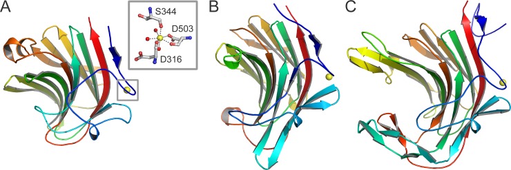
Fig 2. β-Sandwich domain of Xyl in comparison with other enzymes.
The C-terminal β-sandwich domain of Xyl (residues 310–511; A) is similar to that of Bacillus (1,3–1,4)- β-glucanase H(A16-M) (PDB 1AYH; B) and P. carrageenovora κ-carrageenase (PDB 1DYP; C). The structures are in about the same orientation and colored in rainbow spectrum from blue at the N-terminus to red at the C-terminus. Inset: Detailed view of the interactions between the C-terminal domain of Xyl and the calcium ion (yellow). A calcium ion has been found at a similar position in the other two enzymes.

