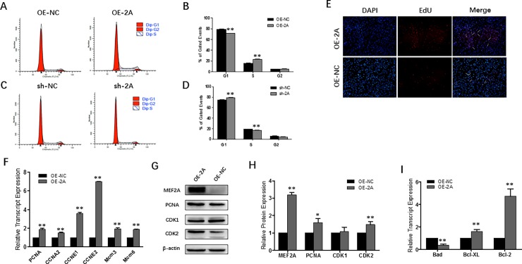
Fig 3. MEF2A promotes myoblast proliferation through triggering cell cycle progression.
(A~B) Flow cytometric measurement of DNA content using propidium iodide (PI) staining in OE-2A/OE-NC treated proliferating myoblast. (C~D) Flow cytometric measurement of DNA content using propidium iodide (PI) staining in sh-2A/sh-NC treated proliferating myoblast. (E) Images of the EdU assay: DAPI staining is shown in blue and EdU staining is in red (OLYMPUS IX71 100×). (F) Relative mRNA expression of cell cycle genes: PCNA, CCNA2, CCNE1, CCNE2, Mcm3 and Mcm6. (G~H) Western blot and protein expression analysis of PCNA, CDK1 and CDK2 showed that MFE2A activated CDK2 expression but not CDK1 expression. (I) Relative mRNA expression of pro-apoptotic gene (Bad) and pro-survival genes (Bcl-2 and Bcl-XL) at early apoptotic stage. Error bars represent s.e.m. *P < 0.05; **P < 0.01.

