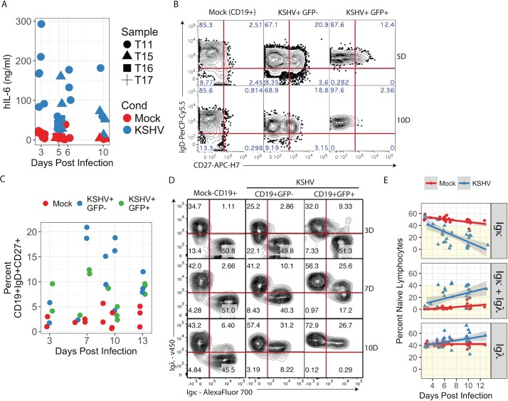
Fig 2. KSHV infection of naïve B lymphocytes recapitulates features of MCD.
(A) Concentrations of human IL-6 in culture supernatants were determined in mock and KSHV-infected naïve B lymphocyte cultures at timepoints between 3 and 10 days post-infection by bead-based immunoassay (BD Cytokine Bead Array). Data represent 9 independent experiments with 4 tonsil specimens. p<0.0001 for aggregate data comparing Mock vs. KSHV. p = 0.003 for Mock vs. KSHV at 5 days post-infection and p = 0.004 for Mock vs. KSHV at 6 days post-infection. Primary naïve B lymphocytes were magnetically sorted from total tonsil lymphocytes and infected with KSHV or Mock infected. At timepoints between 3 and 13 days post-infection 2e5 cells were removed from each culture and analyzed by FCM (B) representative plots gated on single, CD19+ population from a representative experiment at 5 and 10 dpi. (C) Aggregate data for 6 independent experiments from 5 tonsil specimens showing the percent of each subset, which expressed both IgD and CD27 at indicated timepoints post-infection. Additional linear mixed model regression on each independent experiment followed by ANOVA (Type II Wald F tests with Kenward-Roger df) analysis revealed a significant effect of KSHV infection (F = 16.5, p = 0.0005) and both GFP negative (p<0.0001) and GFP positive (p = 0.02) populations were significantly different from Mock based on post-hoc Tukey test. (D) FCM plots gated on CD19+GFP- or CD19+/GFP+ populations from a representative experiment at 3, 7 and 10 dpi. (E) Aggregate data for 11 experiments from 8 individual tonsil specimens showing the percent of the CD19+ population with each light chain phenotype (Igκ+, Igκ+Igλ+ and Igλ) over the timecourse of infection. Linear regression was performed in R software using least means method and gray shading represents a 95% confidence interval. Additional linear mixed model regression on each independent experiment followed by ANOVA (Type II Wald F tests with Kenward-Roger df) analysis revealed significant effects of KSHV infection for each light chain immunophenotype (for Igκ: F = 147.7, p = 2.3E-15; for Igκ+Igλ: F = 52.4, p = 6.3E-9; for Igλ: F = 16.5, p = 0.0002).

