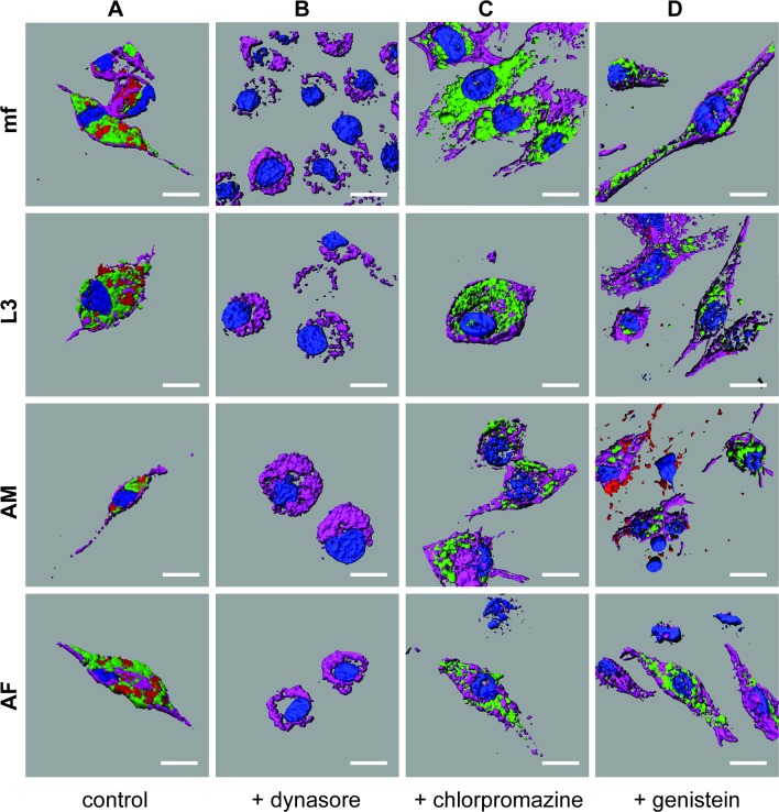
Fig 4. Murine macrophages internalize parasite-derived EV by phagocytosis.
Imaris 3D reconstructed confocal micrographs of murine J774A.1 macrophages. (A) Control macrophages showing internalization of PKH67-labeled EV (green) isolated from microfilaria (mf), L3, adult male (AM) and adult female (AF) worms in parallel with Fluoresbrite Carboxylate Microspheres (red, phagocytosis tracer). Macrophages are counterstained with Hoechst 33342 (nuclei, blue) and phalloidin (muscle, purple). (B) Macrophages treated with labeled EV (green) and microspheres (red) in the presence of 200 μM Dynasore. Absence of green and red indicates internalization of both EV and tracer are blocked. (C) Macrophages treated with labeled EV (green) and Alexa Fluor 555 conjugated transferrin (tracer, red) in the presence of 30 μM Chlorpromazine. Presence of green and absence of red indicates internalization of tracer is blocked but EV is not. (D) Macrophages treated with labeled EV (green) and Alexa Fluor 555 conjugated cholera toxin b (tracer, red) in the presence of 300 μM Genistein. Presence of green and general absence of red indicates internalization of tracer is generally blocked but EV is not. All Imaris images captured at magnification 68X, all scale bars 2 μm.

