Figure 2.
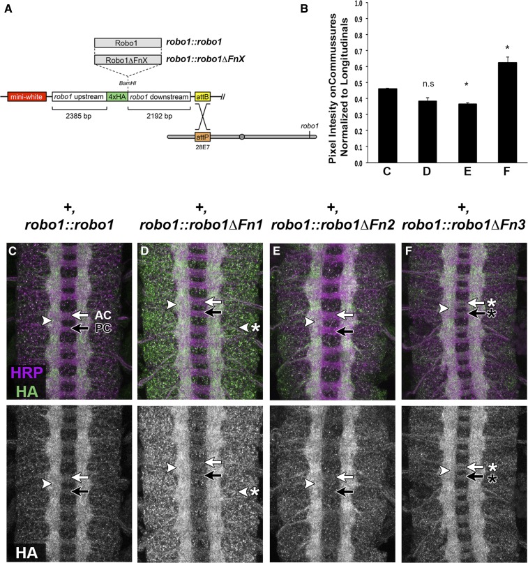
Fn domains 1–3 are not required for axonal localization, and deletion of Fn3 increases Robo1 levels on commissures. (A) Robo1 rescue construct schematic (Brown et al., 2015). HA-tagged robo1 variant cDNAs are inserted between upstream and downstream flanking sequences, which reproduce robo1’s endogenous expression pattern. All transgenes are inserted at the same landing site to ensure equivalent expression levels (cytological position 28E7). (B) Average pixel intensity of anti-HA staining on commissural axons normalized to longitudinal axons for the genotypes shown in (C–F). Pixel intensity was measured for commissural axons at five locations per embryo and normalized to pixel intensity of longitudinal axons from the same segment. Normalized commissural expression levels are shown, averaged over three embryos for each genotype. Each variant was compared to +, robo1::robo1 embryos (C) by a Student’s t-test, with a Bonferroni correction for multiple comparisons. We detect a statistically significant increase in relative expression levels on commissural axons in embryos expressing Robo1ΔFn3 compared to embryos expressing full-length Robo1 (* P < 0.01). (C–F) Stage 16 embryos stained with anti-HA (green) and anti-HRP (magenta) (top), and HA alone (bottom). All transgenic receptors are properly localized on longitudinal axons (arrowhead) and cleared from commissures (arrows), with the exception of Robo1ΔFn3, which is present on commissures (F, arrow with asterisk). Robo1ΔFn1 expression is elevated within cell bodies compared to other transgenes (D, arrowhead with asterisk). AC, anterior commissure; Fn, fibronectin type-III repeat; n.s, not significant; PC, posterior commissure.

