. 2018 Apr 26;7:e33670. doi: 10.7554/eLife.33670

Figure 4. Lower capillary density in aged mice is predicted by obstruction and pruning rates.

(A) Representative confocal images of Evans blue filled blood vessels in somatosensory cortex of young (3–4 month) and aged (15–18 month) mice. Scale bar 200 µm. (B) Box and whisker plot of capillary density (+denotes mean) across all cortical layers for 3–4 and 15–18 month old Tek-GFP mice (n = 6–7 mice; unpaired t(11)=2.47, p=0.03). Error bars are S.E.M. (C) Box and whisker plot of normalized cortical capillary density for 3–4 or 15–18 month old Tek-GFP mice (n(3-4month)=6, n(16-18month)=7 mice, unpaired t(11)=2.47, p=0.03). Error bars are S.E.M. (D) Predicted capillary loss over time (blue line) based on measured rates of spontaneous obstructions and pruning (see Materials and methods, Figure 4—figure supplement 1). Red data points and mean (±SEM) represent measured capillary loss in 15–18 month old aged mice normalized to 3–4 month old mice. Predicted capillary loss closely matched experimentally measured loss in aged mice (unpaired t-test, p=0.52).

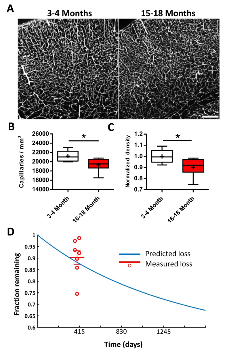
Figure 4.

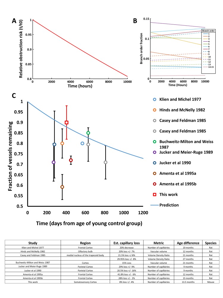
Figure 4—figure supplement 1. Modeling capillary loss over time.

Figure 4—figure supplement 1.

We modeled a hypothetical set of 100,000 capillaries, applying the proportional risk of obstruction (by branch order) and global risk (30%) of subsequent pruning every 2 hr. (A) Plot showing the relative risk of capillary obstruction decreased over time as at-risk capillaries were lost, leaving a higher proportion of less frequently obstructed capillaries (usually of higher branch orders). Note that this ignores possible changes in branch order due to pruning as capillaries were modeled independently with fixed branch orders. (B) Relative fraction of each capillary branch order over time. Over 10,000 hr the fraction of frequently obstructed capillaries were reduced while the number of rarely obstructed capillaries stayed constant and thus comprised a greater fraction over time. (C) Comparing our predicted and experimentally derived estimate of capillary loss to previous aging studies in rodents. Note that some previous studies used histochemical markers to label vasculature such as alkaline-phosphatase, which might decrease in aged animals’ independent of actual vessel less, leading to over-estimates of capillary loss. Data shown was calculated from published numbers in each study, ±S.D. when available.