
Fig. 1.
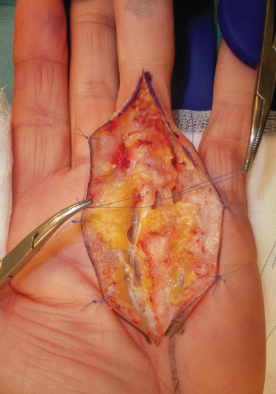
An intraoperative picture showing the radial spiral cord with a serpentine neurovascular bundle, deviated to the ulnar side, around which a suture loop has been passed for identification. The ulnar neurovascular bundle is also shown with a suture loop.