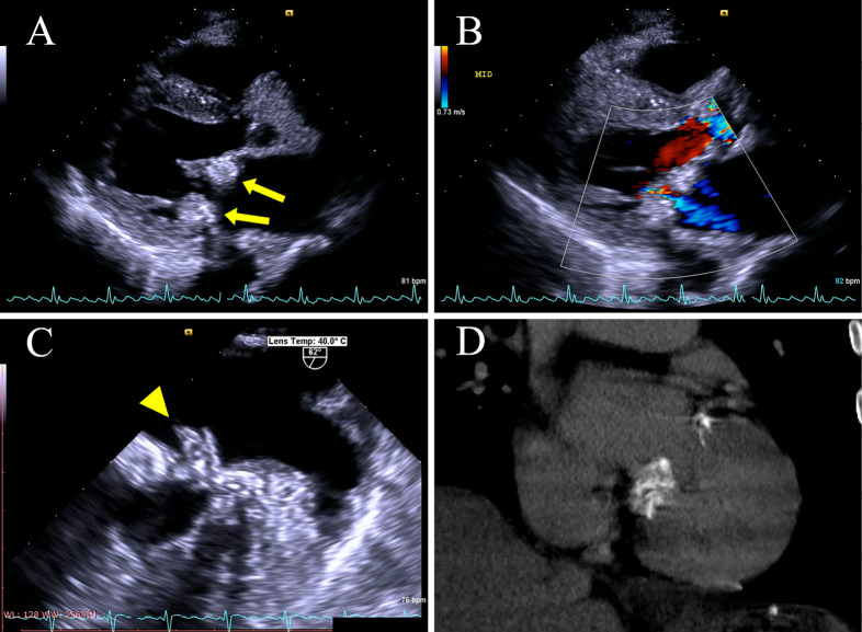
Figure 2.
Transthoracic echocardiography with a long-axis parasternal view after admission (A) shows advanced CCMA progressing on both the posterior and anterior annulus of the mitral valve (yellow arrow). The features of CCMA, such as central echolucency resembling liquefaction, are clearer than they were in the previous test. Color Doppler (B) shows mild regurgitation of the mitral valve, similar to the previous echocardiogram. Transesophageal echocardiography (C) shows vegetation attached to the base of the posterior mitral valve leaflet (P2) (yellow arrowhead). Non-contrast computed tomography (D) confirms the presence of diffuse calcification of the mitral valve.

