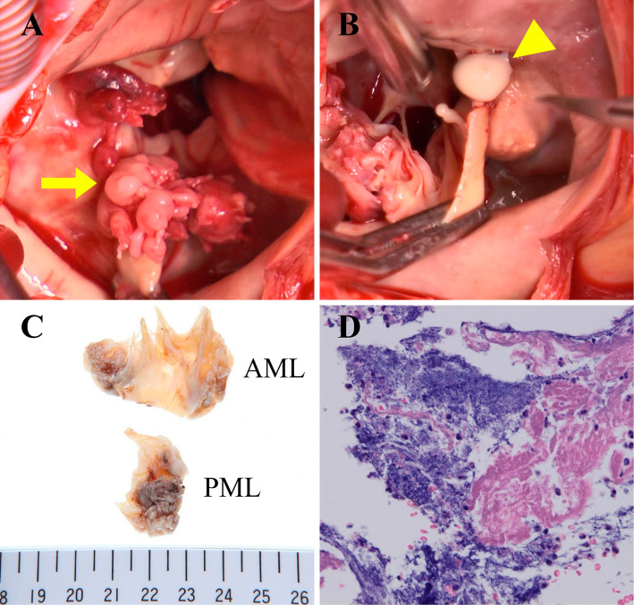
Figure 3.
An intraoperative view (A) shows the vegetation of bacterial endocarditis attached to the base of the posterior mitral valve leaflet (P2). Caseous calcification covers the entire circumference of the mitral annulus. A white pasty material flows out from the incised mass of the mitral annulus (B). The examination of tissues of the mitral valve leaflet (C and D) reveals vegetation of infective endocarditis with neutrophilic infiltration, vascularization and bleeding.

